1) Cell clusters in intervertebral disc degeneration: an attempted repair mechanism aborted via apoptosis Lama, P., Tiwari, J., Mutreja, P., ...Adams, M.A., Le Maitre, C. Anatomy and Cell Biology, 2023, 56(3), pp. 382–393
2) Computational Image Analysis of Painful and Pain-Free Intervertebral Disc. Tiwari, J., Sharma, S.R., Chauhan, S., Adams, M., Lama, P. Lecture Notes in Computational Vision and Biomechanics, Artificial Intelligence on Medical Data, 2023, 37, pp. 373–386
|
Dr. Polly Lama attended and presented oral paper at the International Society for the Study of Spine (ISSLS), Melbourne, Australis held on 01/05/23 to 05/05/23.
1) Title " Machine Learning Based Image Analysis Tool for Hybrid Visualization of Macroscopic - Microscopic Degenerative Changes in Intervertebral Disc"
2) Title "Biological Significance of Fragmented SLRF's Aggrecan and Fibronectin in Overloaded Intervertebral Disc"
3) Presented: Invited Lecture on "Empowering the 21st century higher education outreach in the Medical Science Programme" at the International Hybrid Conference on Outcome Based Education (OBE-2023) held at Mahatma Gandhi University, Kottayam, Kerela on10th to 12th February 2023
|
1) Continent Wise Intersectional Analysis on Ageing. Lama, P, 2023. The Ageing Population: Impact analysis on societal and healthcare cost. In: Lama, P. (eds), Springer Nature, Singapore, 2023, 1, pp.1-34.
2) The Economic Impact of Ageing on Healthcare. Lama, P, 2023. The Ageing Population: Impact analysis on societal and healthcare cost. In: Lama, P. (eds), Springer Nature, Singapore, 2023, 1, pp. 69-81.
3) Future Investment Needs for the Aging Community. Lama, P, Tamang, B, Bhutia C S, 2023. The Ageing Population: Impact analysis on societal and healthcare cost. In: Lama, P. (eds), Springer Nature, Singapore, 2023, 1, pp. 83-90.
4) Variabilities in Ageing Trends: Questions and Concluding Remarks. Lama, P, 2023. In: Lama, P. (eds) The Ageing Population: Impact analysis on societal and healthcare cost. In: Lama, P. (eds), Springer Nature, Singapore, 2023, 1, pp. 91-94
5) Editorial Lama P (2023). The Ageing Population: Impact analysis on societal and healthcare cost. In: Lama, P. (1st edition), Springer Nature, Singapore, 2023, 1, pp.1-118.
6) Lama et al., Computational Image Analysis of Painful and Pain-Free Intervertebral., Springer Nature, Artificial Intelligence in Medical Data, 2023, 37, pp. 373–386.
Link:https://www.springerprofessional.de/computational-image-analysis-of-painful-and-pain-free-
interverte/23298842
|
Dr. Rohit Kr. Sarda : Dolphee Khurana1*, Ankitha Suresh1*, Raghavendra Nayak2,
Manjunath Shetty3, Rohit Kumar Sarda4, Jonathan C Knowles5,6,7 ,Hae-Won Kim6,7,8,9, Rajendra K Singh6,7,8 and Bhisham Narayan Singh1
“Biosubstitutes for dural closure: Unveiling research, application, and future prospects of dura mater alternatives” Journal of Tissue Engineering Volume 15: 1 –26 © The Author(s) 2024
Article reuse guidelines:
sagepub.com/journals-permissions
|
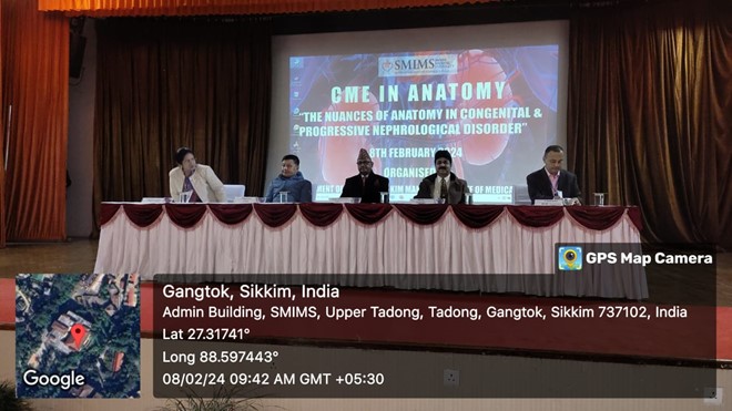
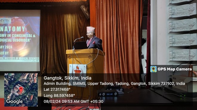
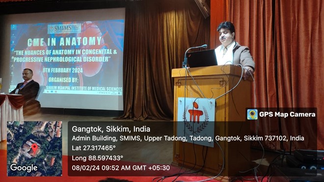
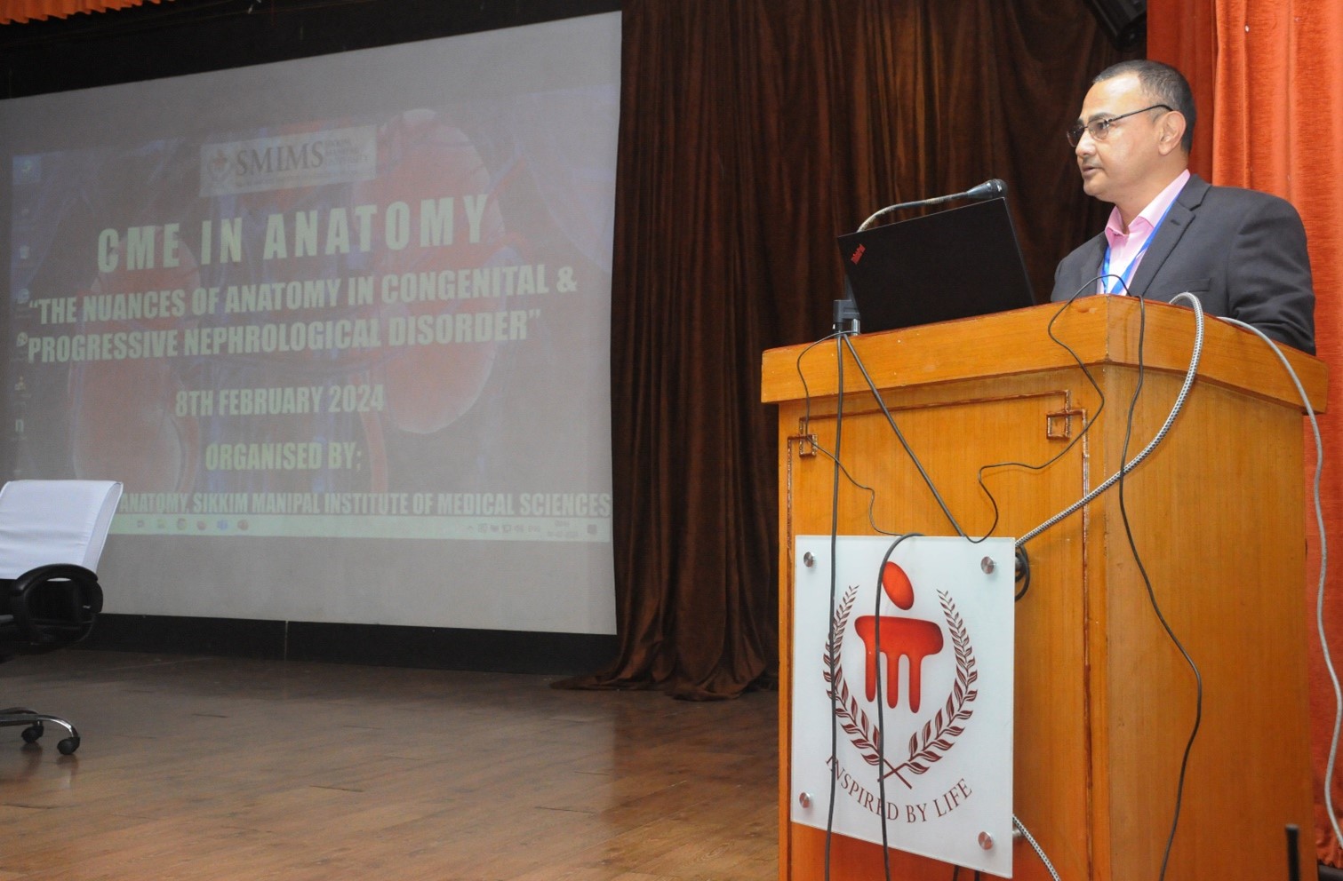
Dr. Poonam Shilal Presented Presented an invited lecture on “Embryological Development of Kidney at CME “The Nuances Of Anatomy In Congenital And Progressive Nephrological Disorder” held at SMU, SMIMS Gangtok. Organized by Department of Anatomy SMIMS.
On 8th February 2024.
|
|
Jerina Tewari, Siddhi Raj Sharma, Sukirti Chauhan, Mike Adams, and Polly Lama " Computational Image Analysis of Painful and Pain - Free Intervertebral Disc Journal of Springer Nature Artificial Intelligence on Medical Data, LNCVB on Aug 2022;vol 37:pg 373 -386. https://doi.org/10.1007/978-981-19-0151-5_31.
Nov 2022 (online first) January 2023 (Print)
|
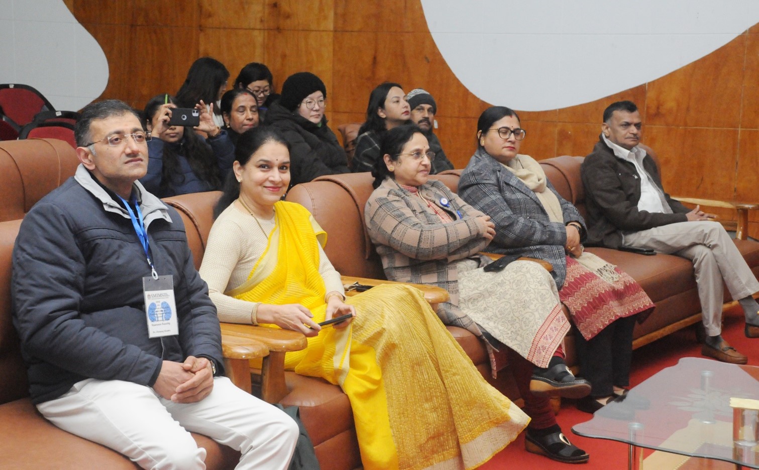
1).Dr. Binod Kr. Tamang, Dr. Polly Lama, Dr. Kalpana Chettri, Dr. Poonam Shilal, Dr. Karma Lakhi Bhutia, Dr. Rohit Kr. Sarda, Jerina Tewari attended the CME “The Nuances Of Anatomy In Congenital And Progressive Nephrological Disorder” held at SMU, SMIMS Gangtok. Organized by Department of Anatomy SMIMS. O 08th February 2024
|
|